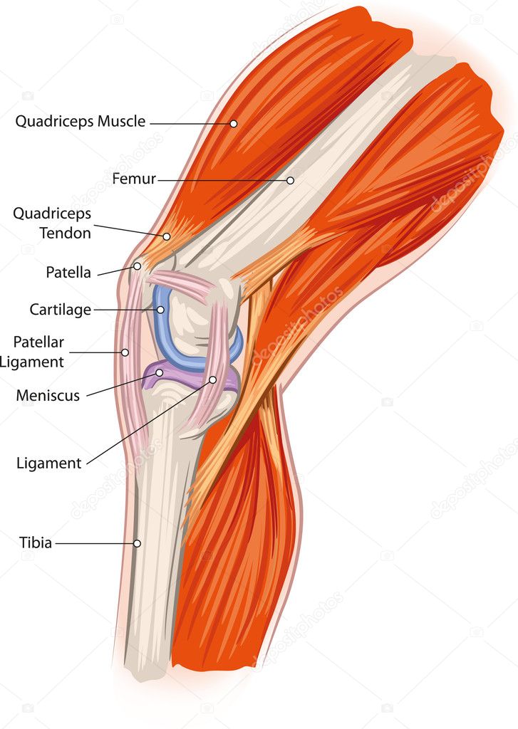
The Knee Joint Anatomy Chart 1001488 Knee Anatomy Poster Biology Diagrams Learn about the knee joint, a hinge type synovial joint formed by the patella, femur and tibia. Explore its articulating surfaces, ligaments, menisci, bursae, neurovascular supply and common injuries.

Learn about the structure and function of the knee joint, a synovial hinge joint formed by the femur, tibia, and patella. See 3D images and diagrams of the bones, cartilage, ligaments, and bursae that support and protect the knee. Learn about the structure and function of the knee joint, the largest and most complex synovial joint in the human body. The knee joint consists of three bones, two menisci, four ligaments, a joint capsule, and various muscles and bursae.
Knee Anatomy: Bones, Muscles, Tendons, and Ligaments Biology Diagrams
Learn about the structure and function of the knee joint, the largest and most easily injured joint in the body. Find out how cartilage, ligaments, muscles, and other tissues work together and what conditions can affect them.

Learn about the knee joint, a complex hinge joint composed of two articulations; the tibiofemoral and patellofemoral joints. Find out the articular surfaces, ligaments, menisci, innervation, blood supply and movements of the knee joint. The accessory bones of the knee are rare anatomical variations that may develop within the soft tissues of the joint. The os fabella is a small sesamoid bone found in the lateral head of the gastrocnemius which can become symptomatic. It is the most common accessory bone of the knee. The cyamella is another sesamoid bone, located in the tendon of the popliteus muscle.
Knee Joint Anatomy: Bones, Ligaments, Muscles, Tendons, Function Biology Diagrams
Learn about the knee joint, one of the largest and most complex joints in the body, composed of four bones, two menisci, four bursae, and various ligaments and muscles. The knee joint allows for flexion and extension, and is stabilized by the menisci, ligaments, and synovial fluid. Learn about the eight components of knee joint anatomy, how they work together and what can go wrong. Find out how to prevent and treat common knee problems such as cartilage, ligament, tendon and bursa injuries.
